
")
")
Analysers
The flow cytometry platform is composed of two analyzers and a cell sorter.
These analyzers allow the multiparametric study of particles in suspension in a flow (biotic or abiotic). Concerning the aquatic microbial ecosystem, they allow its study at the scale of the individual cell. The abundances of the different functional groups can thus be determined (viruses, bacteria, phytoplankton, heterotrophic flagellates) thanks to the differentiation allowed by the measurement of fluorescence intensities for each of the cells passing in front of the lasers, and light scattering at 180° (FSC) and 90° (SSC) which are relative indicators of cell size and internal complexity of the cells. Cells in suspension, living or not, are labeled by excitable fluorophores at the wavelengths available on our instruments (ex: heterotrophic bacteria) or autofluorescent (ex: photosynthetic organisms) and thus detected without labeling. We work mainly on the structure of microbial communities in marine and freshwater as well as on cell cultures.
FACS Canto 2, BD Biosciences
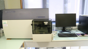
The BD FACS Canto II is equipped with three excitation lasers at 405, 488 and 633 nm and 8 channels of collection of the emitted photons. These 8 channels collect the emitted photons from the excitation of fluorophores (induced or natural) and transform them into electrons thanks to photomultipliers (PMT).
Diva6 is commonly used to work on the datas under Windows XP operating system.
Cytoflex, Beckman Coulter
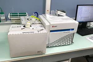
The Beckman Coulter Cytoflex, also equipped with the same 3 excitation colors and as many recovery channels for the emitted photons. It is however, in addition equipped with an SSCv linked to the violet laser dedicated to the study of small particles. The detectors of this instrument are avalanche photodiodes (APD) more sensitive and less noisy than the PMTs.
CytExpert is commonly used to work on the datas under Windows 7 operating system.
Instruments are available through Open Iris.
Oceanographic application:
Flow cytometry allows a good understanding of the complex structure of phytoplankton community. Phytoplankton community is mainly composed of unicellular algae ‹3µm. This cytometric technique enable the seasonal monitoring of the phytoplankton evolution in aquatic ecosystems. This single-cell cellular approach of the ecosystems led to new shade on oceanographic functioning, specifically in biogeochemical studies of the ocean.
New, very small and very abundant microalgae species have been discovered through flow cytometry analyses:Prochlorococcus sp. (Chisholm et al. 1988) or Ostreococcus tauri (Courties et al. 1994). Their primordial role in the trophic food web functioning is mainly studied through flow cytometer & cell sorter, sometimes shipped on marine vessel.
Here you have an example of cytogram that show phytoplankton populations where each one of the cells are identified by scatters & fluorescences.
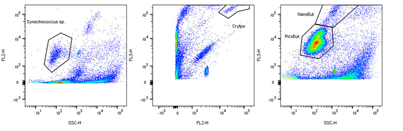 Phytoplankton analysis workflow © David Pecqueur / OOB
The BioPIC platform is notably referent for pico- and nanoplankton analyses of the SOMLIT observation network (INSU-CSOA) which includes more and more observation stations (12 to date). We also collaborate with national and international universities and some private companies on specific projects.
The Cell sorter
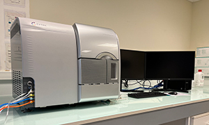
The cell sorter is an AURORA CS (CYTEK) with three lasers of 405, 488 and 640 nm. It is also equipped with APDs but includes 38 allowing to identify precisely the emission spectrum of the detected populations (e.g. phytoplankton). It allows sorting in tubes (1.5ml, 5ml, 15ml) with 2 to 6 sorting ways or in plates (96 wells). It allows in particular the purification of contaminated cultures or the isolation of particular microbial groups for genetic analysis.
![]() White paper: Characterizing Marine Phytoplankton with Full Spectrum Profiling™ Technology
White paper: Characterizing Marine Phytoplankton with Full Spectrum Profiling™ Technology
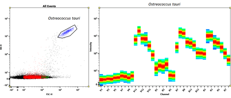 Full spectrum of Ostreococcus tauri marin model © David Pecqueur / OOB
Marine bacteriology application:
We use green dye Syber Green I to count marine bacteria (millions/mL). These bacteria use to show 2 or 3 populations weakly fluorescent (LNA) or highly fluorescent (HNA) and this fluorescence is linked to their activity.
Bacteria cell sorting, identified through green fluorescence and scatter, associated to specific metabolisms analyses or characterized in molecular biology, lead to specify these activities and their diversity still mainly unknown in aquatic ecosystems.