
")
")
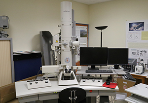
Electron microscopy is a source of innovative studies for marine models such as viruses, bacteria, unicellular algae, marine metazoans. The Oceanological Observatory of Banyuls-sur-Mer has a transmission electron microscope: Hitachi 7500 equipped with a high-resolution camera, AMT advantage.
The department also has a sample preparation laboratory, a room equipped with 2 Ultra microtomes: a Leica, an LKB and cryo-sample preparation instruments: Leica .EM AFS2 and GP.
Example of application on marine model studied in the observatory:
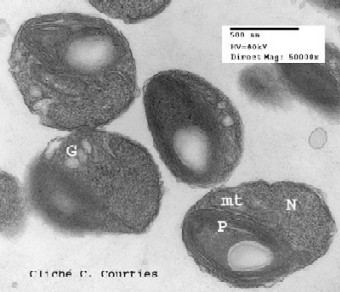 Ostreococcus tauri © Claude Courties / OOB
Ostreococcus tauri is the smallest free eukaryote known. It is a 1μm green alga with a basic structure consisting of a nucleus, a chloroplast, a mitochondria and a golgi apparatus (Courties et al., Nature 1994). Discovered in the pond of Thau where it abounds, it is also present in all the oceans. It has become a study model for cell biology and marine ecology.